Mitosis
he Greek mitos meaning "filament" (referring to the appearance of chromosomes microscopy), the mitosis referschromosomal events of cell division . This is a duplicate "non-sexual" (unlike meiosis ).It is the division of a mother cell into two daughter cells.
It also refers to a very specific stage of the life cycle of cellseukaryotes , called " cell cycle ", which is the stage of separation of eachchromosome from the mother cell and their equal distribution in each of the two daughter cells. Thus, each "core son" receives a complete copy of the genome of the organism "mother." The DNA is replicated by DNA polymerase when it is in the form of chromatin (equivalent to a chromosome place) at the interphase of the cell cycle.
The cell cycle is divided into several phases:
- phase G 1 , early growth phase (the longest)
- S phase during which the genetic material is replicated ,
- Phase G 2 , which is the second phase of cell growth and,
- M phase, that of mitosis itself,
- there is a quiescent phase known which corresponds to the output of the cycle phase G 0 , it usually occurs in G1.
Phases G 1 , S and G 2 are the interphase .
Mechanisms of mitosis are very similar in most eukaryotes , with only minor variations. The prokaryotes have no nucleus and only have one chromosome without centromere , they do not divide by mitosis itself, but split binary, tertiary, multiple, or by budding.
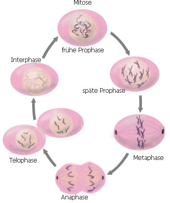
Phases of mitosis
Mitosis is a continuous phenomenon, but to facilitate the understanding of its development, the biologists described four characteristic steps of mitosis arethe prophase , the metaphase , the anaphase and telophase . Mitosis lasts between 1 and 4 hours
Interphase

The interphase is the cycle period cell before mitosis is characterized by an increase in cell volume, the cell transcribes its genes and chromosomes are replicated. It is therefore not strictly part of mitosis. Chromosomes are in the form of compact filaments: chromatin. It is during this phase that the DNA replication takes place (each chromosome doubles, two chromatids ). It can be divided into several phases.
- The phase G 1 (Gap 1 in English, gap = space for the space between mitosis and S phase) during which the cell grows and performs the functions for which it is genetically programmed: protein synthesis, etc. .This phase determines the final size of the daughter cells from mitosis.
- The S phase (for Synthesis of new molecule of DNA ) in which the chromosomal material (currently in the form of chromatin) is doubled by duplication. Each filament of chromatin is split into two filaments remain stuck in a sort of cross (the cross will, by compacting / rolling / condensation usually called chromosome , that is to say two chromatids bonded their centromeres).
- The phase G 2 (Gap 2) wherein the cell behaves as in phase G
Prophase
Prophase is organized into filaments more net.During this phase, the genetic material ( DNA ), which is normally present in the nucleus in the form of chromatin condenses into highly ordered structures and individualized called chromosomes .Indeed, proteins called histone H1 are attached from one side of the DNA. However, during prophase, these are phosphorylated histone H1 (by MPF) which causes an increased winding of DNA that seems to "condense". Thenucleolus disintegrates. Since the genetic material has been duplicated before mitosis, there are two identical copies of each genotype cell . During this phase, the chromosomes are composed of two chromatids sisters both wearing the same genetic information. They each also contain a DNA element called the centromere , which plays an important role in chromosome segregation.Both chromatids of the same chromosome are joined at the centromere region.A protein called cohesin acts as glue and unites the two chromatids of a chromosome.
The second organelle important prophase is the centrosome , composed initially of two centrioles in animal cells (except those who have lost their ability mitotic, as most of the nerve cells that contain none, and cancer cells that may contain more; centrosomes plant cells do not contain one). As the chromosomes, the centrosome is duplicated before the start of the prophase(centrioles in 4). 4 centrioles separate during prophase , forming two centrosomes that migrate to each pole of the cell. The cytoskeleton ofmicrotubules is reorganized to form the mitotic spindle , bipolar structure that extends between the two centrosomes but remains outside the nucleus. By the growth of microtubules , the mitotic spindle elongates, which stretches the cell nucleus .
We can represent the microtubules as poles or rails in the cell. Some cellseukaryotes , including plant cells, are devoid of centriole .
Metaphase
Second phase of mitosis after prophase, the gathering of condensed chromosomes at the equator of the cell to form the equatorial plate .The stress on each kinetochore of a chromosome is gradually balanced and they are aligned in a plane located midway between two poles. It is observed that the chromosomes are aligned along their centromere.
Anaphase
he anaphase phase is very rapid meiosis and mitosis where the chromatids separate and migrate to opposite poles of the cell. The son chromosome on which hung the centromeres of cells detach and chromatids move to each pole of the cell.
During this phase, following a specific signal which corresponds to a 10-fold increase in the concentration of intracellular calcium and inactivation of MPF (cyclin B proteolysis MPF), the chromatids abruptly separate sisters. They are then "learned" by microtubules towards the pole to which they are attached.Chromatids migrate rapidly at a speed of about 1 micrometer / min. There are two categories of movement: anaphase A and anaphase B.

During anaphase A chromatids in reality move poleward on kinetochore microtubules that shorten as they depolymerize at their end + As the progress of the kinetochore.Indeed, the kinetochores not only allow "secure" a chromatid microtubule, but also to be transported along microtubules. At kinetochores found "engines" molecular (such as dynein ) using the ATP that can pull the chromatids along microtubules that they remain fixed.
During anaphase B, the polar microtubules lengthen, and mitotic spindle poles move apart from one another bringing with them the chromatids
Telophase

The term "telophase" derives from the Greek word "telos" meaning "end". This is the 4 thphase of mitosis.
During this period:
- polar microtubules will persist in their end + to form interzonal microtubules which disappear when the highest phase terminal telophase, cytokinesis, which corresponds to the terminal division of two daughter cells.
- The kinetochore microtubules disappear.
- The chromatid sisters begin to unpack.
- The nuclear envelope and nucleoli begin to reform in métaiose.
Cytokinesis
Still called cytokinesis and cytokinesis , it is after mitosis. During this period, the dividing groove is formed in a plane perpendicular to the axis of the spindle and separates the cell into two. It can actually begin to form in anaphase.Cleavage is due to a contractile ring is composed mainly of actin and myosin(myosin II). This constriction is centripetally. The cleavage furrow tightens up form an intermediate body, forming a narrow passage between the two daughter cells and which contains the rest of the mitotic spindle. It will eventually disappear completely and the two daughter cells will separate completely.Moreover, the nuclear envelope and nucleoli to recover and finish the interphase radial arrangement of microtubules nucleated by the centrosome is reformed.
In the plant cell , cytokinesis is very different from the presence of a rigid wall (divided into a primary wall, cellulose, and a wall primitive, pectinic, the whole forming a wall pecto-cellulose). It is achieved through the construction of a new wall phragmoplast simply called intermediate body between the two daughter cells. This new wall is developing centrifugal of Golgi vesicles containingpropectine accumulate the cell center towards the periphery, and these vesicles fuse to form the phragmoplast which adjoins the primary wall of the parent cell, causing it to division into two daughter cells. The primary wall and the membrane of the two daughter cells then are reformed at the separation and differentiates into phragmoplast middle lamella , or primitive wall.
Consequences of mistakes
Of course, errors can occur during the formation of new cells: the process can indeed wrong place. And when these errors occur during the first mitotic cell division of a zygote , they may be particularly harmful.
Examples of mitotic errors:
1. Phenomenon of nondisjunction : a chromosome does not separate during anaphase. One daughter cell will receive two homologous chromosomes and the other will receive none. One of the daughter cells will then have a trisomy and the other a monosomy , which are cases of aneuploidy .
2. Deletion, translocation, inversion, duplication chromosomiale:
Mitosis is a traumatic process. The cell undergoes significant changes in its ultrastructure, its organelles disintegrate and reform after several hours, and chromosomes are constantly moved by microtubules . Occasionally, chromosomes can be damaged. An arm of the chromosome can be broken and the fragment is lost, causing a deletion . The fragment may be incorrectly attached to another chromosome non-homologous, which causes translocation. It can be reattached to the original chromosome, but in reverse, causinginversion . Or it may be wrongly considered as a separate chromosome, causing then a chromosomal duplication . The effect of these anomalies depends on the specific nature of the error. Sometimes there will be no consequences , other times it can induce cancer , or even cause the death of the body.
Meiosis and mitosis
Mitosis and meiosis differ in a number of points, but also exhibit similarities (chromosome separation mechanisms, etc..). Mitosis is asexual reproduction of cells, while meiosis is a prelude to sexual reproduction. By each parent meiosis produces gametes different destined to meet. Many types of cells are capable of mitosis but only those of the reproductive organs, the gonads (ovaries and testes ) produce meiosis. From a cell at the end of mitosis there are two genetically identical cells while at the end of meiotic cells there are four genetically different most often and therefore unique.
Plant mitosis
The main differences between mitosis and plant mitosis are animal centrioles in the absence of plants (except in some algae and gametes), the presence of a wall which leads to a cytokinesis particular, its role in the postembryonic development and its hormonal regulation. Plant mitosis is still poorly understood, including how the spindle may occur in the absence of centrioles and centrosomes (but at each pole early prophase was called cytoplasmic condensation polar cap that emits radiation which will form the end of prophase the mitotic spindle). Therefore the difference is that for the animal cell in mitosis at the poles were asters from centrioles and the plant cell is a polar caps from the condensation of the cytoplasm. However the events of mitosis are strongly linked to cytoskeletal rearrangements.
Cytokinesis The separation of daughter cells produced by forming a new wall pectocellulosique on the equatorial plane of the cell. This plan is determined by the location of certain proteins at the beginning of mitosis. At the end of telophase microtubules form at equatorial plate, the phragmoplast . Membrane vesicles from the Golgi apparatus and precursors of wall components come join them.
Role in the development In unicellular organisms the availability of nutrients in the medium is the main factor regulator of mitosis that actually depends on the size of the cell. In multicellular organisms divisions occur only in meristems, and the meristematic cells depend for regulating the cell cycle (as for the supply of nutrients) signals generated by somatic cells (phase G 0 , that is ie quiescent, which do not divide): it is social control. The formation of tissues and organs occurs at the level of accumulation of meristem cells (mérèse).
The mérèse taking place only in meristems, if a somatic cell is damaged or destroyed it is not replaced, unlike what happens in the animal kingdom. So that the plants do not have an organizational plan as strict as that of animals, there is formation of new organs and senescence old. Another difference is that in plants the apoptosis is not significant in the formation of organs.
Hormonal regulation of differentiation signal is given to immature cells in the mature cells. Signals can be non-peptide hormones ( auxin , cytokinins ,ethylene , abscisic acid , brassinosteroids ), lipo-oligosaccharides ( Nod factor ) peptide ( systemin ). The hormone response is variable depending on the tissue. It acts via MAPK genes (MAPK cascades), triggering the accumulation of cyclin required for entry into S phase
Auxin and cytokinins play a major role in concert mitosis. The exogenous auxin is required for meristem that can be self-sufficient cytokinins. If either is missing hormones levels sufficient mitosis does not occur. Auxin activates the expression of genes SAUR (response 2-5 min) and AUX / IAA (5-60min response). It acts mainly on secondary meristems (cambium mainly).Cytokinins stimulate chromosome segregation and cytokinesis, causing the accumulation of cyclins and activate cdc25 phosphatase that activates cyclin kinase cdc2 by dephosphorylation of tyrosine 15. They are necessary for the initiation of cell cycle progression as his.
ABA inhibits mitosis in response to water stress by inducing the synthesis of ICK inhibitor of cyclin-cdk in meristematic tissues. Brassinosteroids and gibberellins promote mitosis. Gibberellins stimulate the proliferation of intercalary meristems (monocots) and cortical and epidermal tissues insensitive to auxin by increasing the expression of histone H3 and cyclin 1.
Nod factor initiates root nodulation in the presence of bacteria Rhizobium.
Nod factor initiates root nodulation in the presence of bacteria Rhizobium.
In response to a stress reduces plant growth organs slowing the cell cycle which reduces the rate of mitosis and the final size of new bodies (they contain fewer cells). This effect is greater in the roots than in the leaves. Response to water and salt stress occurs through the ABA increases the expression of CDKA ICK1 that interacts with and inhibits the activity of histone H1 kinase.More cyclin kinase cdc2 is deactivated by phosphorylation (phosphorylation of cdc2 is considered a major reduction of cell division in response to stress).Another messenger of stress is jasmonate , involved in the response to injury, disease and synthesis of plant cell walls that neutralizes the activity of cytokinins and inhibits cell division. The sensitivity of the cells depends on the jasmonate-cycle phase (largest in G 1 ).
Environmental signals affect growth and cell division. This is a form of plant adaptation to environmental changes. Quiescent cells (G 0 ) may occasionally under the influence of hormonal factors (auxin), nutritional or environmental (light) iron phase G 1 to initiate a cycle of division. This holding capacity mitotic quiescent cells achieves environmental resources (light and minerals).
Maintaining three genomes In addition to the plant nuclear genome must replicate their genomes mitochondria and chloroplasts. Replication of these genomes occurs only in meristems and organs paramount. When the cell is rapidly dividing the number of genomes per organelle greatly increases. When the speed of division slows replication ceases genomes and the number of organelles by cell division increases until there is only one or two genomes by organelle.